Authorized status (The lawful standing is an assumption and isn't a legal summary. Google hasn't done a authorized Investigation and helps make no representation as for the accuracy of your position stated.)
Fluorescence microscopy is extremely sensitive, certain, responsible and extensively used by researchers to look at the localization of molecules in just cells, and of cells in tissues. Fluorescence imaging within reason Light within the sample, which facilitates the visualization of molecules and dynamic procedures in Stay cells.
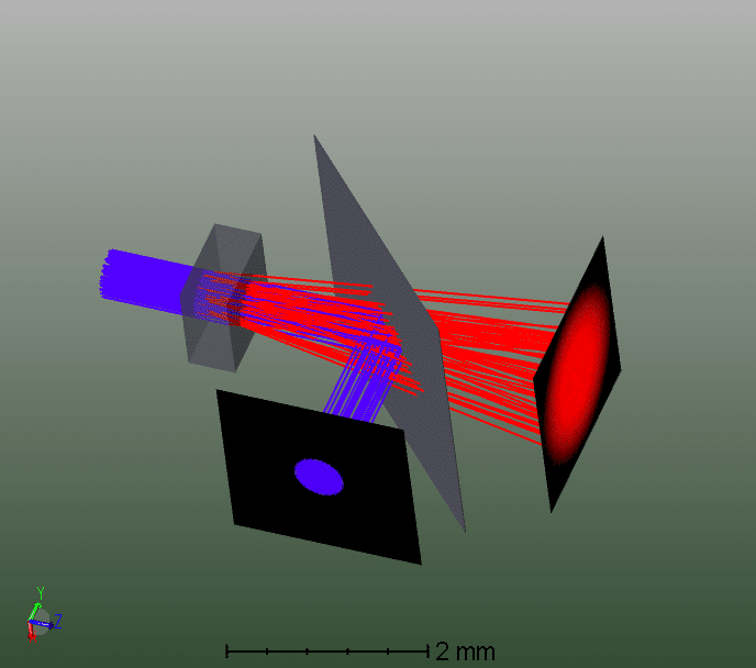
Right here, we created a novel modular microscope which is Charge-productive and suited to imaging diverse low-light-weight luminescence modes. Effects demonstrate this microscope technique characteristics outstanding aberration correction abilities and enhanced graphic resolution, the place bioluminescence, radioluminescence and epifluorescence photos ended up captured and in contrast Using the professional bioluminescence microscope.
The final purpose of this disclosure is to arrive at an exact three-dimensional illustration on the structural attributes from the tissue staying tested depending on multimode optical measurements. To get an accurate, large resolution design in an affordable length of time, the disclosed program begins Using the multimode measurements, makes values with the degree of linear polarization and fluorescence anisotropy as capabilities of wavelength λ.
Γ t o t = Γ r a d + Γ n r a d displaystyle Gamma _ tot =Gamma _ rad +Gamma _ nrad
FIG. 9 displays major see of pores and skin with mole problem for example of tissue with anomaly. Mole has bigger amount of melanin Look at to regular skin for that reason has bigger optical attenuation.
IX(λ) could be the depth of linearly polarized remitted light perpendicular into the input polarization at wavelength λ.
The little silica "bottles" are full of medicine plus a temperature-sensitive material to eliminate malignant cells only in specific parts of your body.
Spatial measurements of intensity as a purpose of wavelength and relative polarization of remitted light are discovered to allow design of three-dimensional useful pictures with the tissue also to extract the location and character of various anomalies, significantly non-malignant and malignant skin lesions.
To make sure agent optical cross sections are very well understood through the UV into the LWIR, quantity built-in measurements of aerosolized agent material at a few key wavelengths is necessary to validate present simulations.
will be the sum of all fees of psyched point out decay. Other premiums of energized condition decay are because of mechanisms besides photon emission and therefore are as a result frequently identified as "non-radiative premiums," which often can include things like:
Therefore, There's been an unmet require for any way of diagnosing melanoma that is certainly connected straight to very well recognized click here physiological parameters, that gives adequate biological plausibility for clinicians, that minimizes the necessity for giant and expensive scientific trials, that gives quantitative 3 dimensional maps of tissue to guidebook remedy, that can provide ample specificity to lessen false positive success and avoidable cure and that substantially gets rid of the masking result of melanin in The natural way darker skin. The present creation gives methods to present these and also other positive aspects.
FIG. twelve is actually a block diagram illustration of the technique for capturing tissue information employing numerous optical modes.
Amongst the anecdotes of navigation by bioluminescence is one recounted via the Apollo thirteen astronaut Jim Lovell, who being a navy pilot experienced identified his way back to his plane carrier USS Shangri-La when his navigation methods unsuccessful.